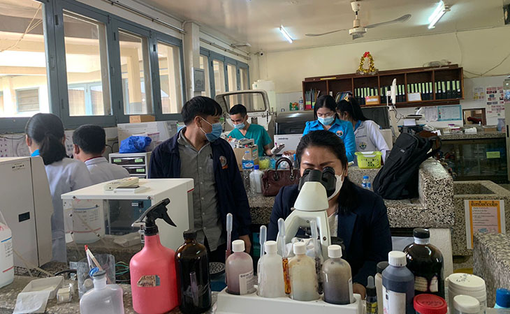
In-service bench training begins in Lao PDR


Lao PDR
March 7, 2022
A major component of the SEALAB project is to provide academic and practical training for topics selected by the laboratories participating in the in-service training program.
An virtual academic training on red cells and white cells pathology and management of inconsistent results was given from November 17-19, 2021 to provincial laboratories.
After receiving the approval from the MoH for the work plan to deliver practical training in the provinces, the Merieux Foundation’s office began the program on February 23-24, 2022 at Oudomxay Provincial Hospital followed by Xayabouly Provincial Hospital on March 3-4, 2022.
The direct evaluation through light microscopy of May-Grünwald-Giemsa (MGG) stained blood smears is still considered the “gold standard” for laboratory identification and count of nucleated blood cells in the blood. This technique can provide additional diagnostic information and should always be used to validate automated methods.
The first bench training given by Ms. Khamphaeng Chanthavong from Mahosot Hospital Microbiology Lab aimed at refreshing the practice of this essential technique at Oudomxay Hospital Laboratory. Mr. Phanthamit Piyadeth, Mr. Bouncher Sisommuang, Ms. Nidsay Saphithack, Ms. Nibdao Souliyanon, and Ms. Phua Wah have been trained to perform May Grunwald-Giemsa stain (MGG), which is used routinely for staining cytological smears. Mrs. Bounleuang Feuang-Inpeng, from Mahosot Hospital Microbiology Laboratory provided a similar training to Mrs. Phetsamone Phanvonsa, Mrs. Vongkhamphone Phetlangsy, Mrs. Phang Soukaserm, Mrs. Phonsavanh Phomphackdy, Mr. Jaithin Saejao and Mrs. Valanud Luanglath.
They reviewed the procedure to prepare MGG staining, preparation, fixing, staining, rinsing the smear and finally, mounting the slide and examining under microscope. They are now familiar with studying cellular morphology, particularly cytoplasm, granules, vacuoles, and basement membrane and with morphological inspection, differential counting of blood cells, erythrocytes, platelets, lymphocytes, monocytes, neutrophils, eosinophils and basophils. They can also make differentiation of normal and abnormal blood cells.
This training will improve support by the lab for the diagnosis of common medical conditions in the hospital.
Peripheral blood smears are the mainstays of diagnosing disorders of blood cells. For example, anemia is very common in Southeast Asia and has many causes, which are classified based on red cell morphology. Recognizing the common white cell and platelet abnormalities is critical to suspect various leukemia and to identify cause of thrombocytopenia.
Additionally to the modules and organization of training courses, the Merieux Foundation has provided necessary reagents and materials, microscope slides, bridges of coloring, May-Grünwald solution and Giemsa stain and smears to show abnormalities of blood count.